
From the moment when the sperm meets the egg through to birth, your baby will go on a complicated journey of development. The first few weeks of the pregnancy are critical and lay the foundation for the birth of a healthy baby. Unfortunately, due to its complexity, this is often the stage of pregnancy where difficulties arise, which may lead to the loss of the baby. Your doctor will assess your pregnancy using blood tests and/or ultrasound if he or she thinks there could be an issue at these early stages. The 5 week ultrasound pictures are usually taken to visualize your baby’s development in the womb, and to assess his or her progress.
How Is Your Baby Developing at 5 Weeks?
Your baby will now be growing rapidly inside your womb, although right now, he or she will appear more like a tadpole than a baby, and will only be the size of a pinhead. The embryo consists of three layers: the outer ectoderm, the middle mesoderm, and finally the inner endoderm, which will go on to form all the baby’s internal organs and tissues. The major organs, including the heart, liver, kidneys, and stomach, are already beginning to take shape, along with the digestive, nervous, and circulatory systems.
The 5 week ultrasound pictures will depend on these development.
What Can You See at the 5 Week Ultrasound?
When you get your ultrasound at 5 weeks, you’ll see the yolk sac and gestational sac. Your baby now will be the size of a sesame seed.
For mothers expecting identical twins, there will be one gestational sac and two yolk sacs; for non-identical twins, there are two gestational sacs and two yolk sacs, with an embryo forming in each.
The gestational sac contains the amniotic fluid, and appears as the black area on your 5 week ultrasound pictures. The yolk sac provides nutrients to the developing embryo, and appears as a small white circle within the gestational sac. It becomes smaller in relation to the baby as pregnancy progresses and the placenta starts to fully form.
Experienced sonographers will be able to find the yolk sac at 5 weeks using transvaginal ultrasound. At this stage, the gestational sac has a diameter of 8-10 mm. The presence of the yolk sac indicates that you have an intrauterine pregnancy, so ectopic pregnancy can be ruled out. However, very occasionally, intra- and extrauterine pregnancies occur simultaneously, and, as with any ectopic pregnancy, this requires urgent medical attention.
5 Week Ultrasound Pictures
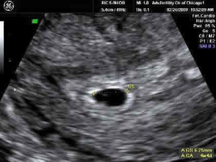
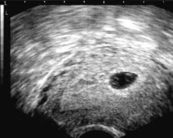
In this transvaginal five week ultrasound image, you can see the gestational sac as the black area on the screen, together with the yolk sac, the small white disc on the left-hand side of the gestational sac. This yolk sac is providing nutrients to the embryo, but the embryo is too small to show up at this stage.
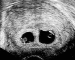
Here, the mother is expecting non-identical twins, as can be seen from the two separate gestational sacs (black areas). The white rings inside the gestational sacs are the yolk sacs. The embryos will have already developed the fetal poles, which are the beginnings of the babies’ cardiac systems.
This is a picture of identical twins, who have developed from the same fertilized egg, and are now seen within the two yolk sacs (white circles) inside the one gestational sac (black area).
This is what one mother sees in her 5 week ultrasound:
Will I Be Able to Hear Heartbeats?
If you have an early ultrasound scan at around 8 weeks pregnant, it may be possible to see and hear your baby’s heart beat for the first time, as the embryo’s heart begins to pulse at about six weeks. At first the baby’s pulse will be between 60 and 90 beats per minute, although the heartrate is not nearly as important as whether there is a heartbeat at all or not.
If you don’t have any scans this early, then the first time you’ll hear the heartbeat will probably be during a regular prenatal care visit. A healthcare professional will use a fetal Doppler to detect the pulse, which may be heard at 10 weeks, although it’s more likely at 12 weeks. The chances of finding the heartbeat depend on the embryo’s position in the womb, the accuracy of your due date, and your own body weight and composition.