
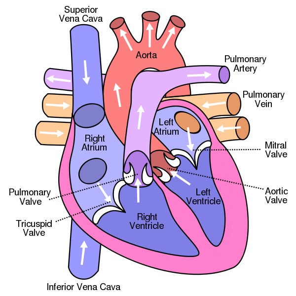
Heart is a vital organ that you cannot live without. The function of heart is quite complex, but you can understand things better through the heart diagram labeled below. It provides information about different chambers of the heart and valves that help transfer blood from one part of your heart to another. Keep reading to learn more about how your heart works.
Four Chambers of the Heart and Blood Circulation
The shape of the human heart is like an upside-down pear, weighing between 7-15 ounces, and is little larger than the size of the fist. It is located between the lungs, in the middle of the chest, behind and slightly to the left of the breast bone. The heart, one of the most significant organs in the human body, is a muscular pump, which pumps blood throughout the body. It beats approximately 72 times per minute, and pumps oxygenated blood to different parts of the body.
Atria and Ventricles
There are four chambers in your heart that are left atrium, right atrium, left ventricle, and right ventricle.
- The upper chambers of your heart are atria, whereas the lower chambers are ventricles.
- Deoxygenated blood enters your heart through the right atria from where the blood moves into the right ventricle first and then to the lungs through the pulmonary artery. Your left atrium receives oxygenated blood from the lungs, which is then pumped into the left ventricle from where it moves into the aorta and then to the different parts of your body.
Valves to Ensure Unidirectional Blood Flow
Your heart has four types of valves with primary function of regulating the blood flow through the heart. Every heart diagram labeledwill clearly show these valves. These valves allow blood flow in one direction only. Different valves perform different functions.
- Tricuspid valve is located between the right ventricle of your heart and the right atrium, and allows the blood to move from the right atrium to the right ventricle.
- Pulmonary valve is between the left pulmonary artery of your heart and its right ventricle. It opens up when the right ventricle contracts and allows the blood to move into the left pulmonary artery.
- Similarly, mitral valve has two cusps and is located in a way that it causes a separation between the left ventricle of your heart and the left atrium of your heart. It pumps the oxygenated blood into the left ventricle when the left atrium contracts.
- Aortic valve separates the aorta from the left ventricle and regulate the flow of blood from the ventricle to the rest of your body.
Blood Vessels
- Blood vessels help transport blood to-and-fro from your heart. These vessels connect other organs in your body to your heart.
- The basic function of these vessels is to take deoxygenated blood from different organs, supply it to the heart, and then take oxygenated blood that comes from the lungs into the heart to the rest of your body.
Blood vessels are more like a network that helps circulate blood throughout your body. There are two types of them: arteries and veins.
- Arteries: These types of blood vessels take oxygen-rich blood from the heart and transport to the capillaries. Arteries are quite tough on the outside but are smooth on the inside. There are three arteries of the heart, including pulmonary artery, aorta, and coronary arteries. Pulmonary artery is the only artery that takes deoxygenated blood from the right side of your heart to your lungs; aorta is the main artery of the heart and transports oxygenated blood to the rest of your body; and coronary arteries are attached to the heart and transfer oxygen-rich blood to your heart muscles.
- Veins: Although they are quite like arteries, they aren't as strong mainly because they don't transport blood at high pressure. The veins receive waste products after the exchange of oxygen and carbon dioxide. Three veins of the heart are pulmonary vein, Venae Cavae, and coronary sinus. Pulmonary vein transfer oxygenated blood to the left side of your heart, venae cavae takes deoxygenated blood back to the heart, and coronary sinus receives deoxygenated blood and transfers it to the right atria.
The Circulation of Blood
Your circulatory system works to transfer oxygenated blood to all the tissues of your body. Your heart pushes blood out when it contracts. It moves in two cycles. In the systemic loop, controlled by the left side of your heart, the blood circulates in your body and supplies oxygen to your organs, tissues, and other structures. In the pulmonary loop, controlled by the right side of your heart, the blood moves to the lungs to release carbon dioxide and get new oxygen.
In the systemic loop, the oxygenated blood comes from the lungs and enters the left atrium, or the upper left chamber of your heart. The chamber presses on the mitral valve once filled with blood and the blood flows into the left ventricle. The blood on the left side of your heart will go into the aorta when the ventricles contract during a heartbeat. The aorta is about an inch wide and supplies oxygen-rich blood to the body's cells. The used blood them moves back and is collected into the two veins, the superior vena cava that receives blood from your upper body, and the inferior vena cava that receives blood from your lower body.
These veins empty into the right atrium of your heart from where your blood moves to the right ventricle through the tricuspid valve. Ventricle contracts and pushes the blood into the pulmonary artery that sends blood to your lungs from where oxygen-rich blood returns to the left ventricle and the process continues.
Exterior of the Human Heart
A heart diagram labeled will provide plenty of information about the structure of your heart, including the wall of your heart. The wall of the heart has three different layers, such as the Myocardium, the Epicardium, and the Endocardium. Here's more about these three layers.
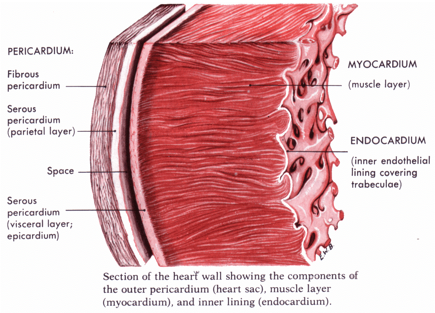
Epicardium
The outermost layer of your heart wall is called the epicardium, which is basically a very thin layer of serous membrane. The membrane provides lubrication and protection to the outer side of your heart, as you can see in heart diagram labeled.
Myocardium
Right beneath epicardium is another relatively thicker layer called myocardium. This muscular middle layer of heart wall contains cardiac muscle tissue. Most of the thickness and mass of your heart wall is made up of the myocardium. The layer is part of the heart that is pumps blood through your body.
Endocardium
Under the myocardium, there is another thin layer called the endocardium. The layer lines the inside of your heart and is usually very smooth. The main role of this smooth, thin layer is to prevent your blood from sticking into the sides of your heart. It also helps prevent the formation of deadly blood clots.
Watch this video to learn more about the structure of human heart.
Different Types of Heart Conditions
Coronary Artery Disease
The arteries that supply blood back to the heart may become clogged due to the buildup of cholesterol plaque. This blockage will cause coronary artery disease. These narrowed arteries may develop a blood clot at any time that leads to a condition called heart attack.
Myocardial Infarction
Also called heart attack, myocardial infarction is the outcome of sudden blockage of a coronary artery. The blockage reduces the amount of oxygen that gets into your heart, which in turn proves fatal for the heart muscle.
Arrhythmia
The condition is characterized by irregular or abnormal heart rhythm caused by changes in the conduction of electrical impulses through your heart. Arrhythmias can be benign or have life-threatening consequences.
Congestive Heart Failure
You develop this condition when your heart becomes too stiff or too weak to pump enough blood through your body. If you're noticing symptoms like shortness of breath and leg swelling, this may indicate congestive heart failure.
Myocarditis
You develop this condition when your heart muscle becomes inflamed. In most cases, this inflammation is the outcome of a viral infection.
Cardiac Arrest
It is a type of heart failure because it refers to the sudden loss of function of your heart.
Sudden Cardiac Death
When someone dies because of cardiac arrest or sudden loss of heart function, it is called sudden cardiac death. The condition is sometimes known as cardiac arrest.
Heart Valve Disease
You have four valves in your heart and issues with any one of them will lead to the development of a heart disease. When not treated early, heart valve disease can lead to the congestive heart failure.
Heart Murmur
When your doctor listens to your heartbeat with the help of stethoscope, he may hear abnormal heart sound. This condition is called heart murmur, which is mostly benign. It may sometimes suggest heart disease as well.