
One of the most frequent shoulder injuries is a rotator cuff tear. Over 2 million people in the United States suffer rotator cuff issues every year and need to have a rotator cuff MRI. It is a major cause of shoulder pain and weakness and accounts for a large amount of missed work, sports and school due to injury. It can also be very disruptive of even simple daily tasks like hair brushing, lifting, and getting dressed for the day.
The rotator cuff tear MRI is a very detailed way of differentiating different types of rotator cuff injuries. It gives very specific images of all the rotator cuff tendons involved and the extent of any damage. It can give doctors needed information on how to treat. Doctors will usually order the MRI if a definitive diagnosis cannot be made on physical examination alone. It also helps eliminate the need for unnecessary rotator cuff repair surgery, but also helps the doctor identify which cases need surgical repair.
Rotator Cuff Tear Overview
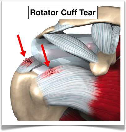 The rotator cuff is a critical part of the shoulder joint. The three bones humerus, scapula, and clavicle are all held together with the help of the rotator cuff. It prevents dislocation of the joint and allows for rotation and lift of the arm. There is also a bursa that lies in the middle of the bone and rotator cuff to give the tendons freedom with arm movement.
A tear to the rotator cuff tendons releases them from the head of the humerus. This can lead to shoulder instability and pain with movement. The condition usually started as a fray in the tendon and then moves on to a full tear. The type of tear is classified into two categories:
- Partial Thickness Tear – A partial thickness tear is just a slight tear, which does not completely go all the way through the tissue.
- Full Thickness Tear – A full thickness tear is a complete tear all the way through the tendons. The tendons may also completely separate from the humerus. This leaves an actual hole.
Rotator Cuff Tear Causes
There are two main causes of rotator cuff tears. These include:
- Attrition – This is a wearing down of the tendons over a period of time from regular usage of the shoulder. The damage is progressive and eventually leads to a tear.
- Injury – Acute trauma to the shoulder leads to a tear in the tendon. This isn’t as common as attrition, but far more painful. These are caused by the shoulder being twisted rapidly and common in people over the age of 40.
How to Know If You Have a Rotator Cuff Tear
The symptoms are caused by inflammation in the tendon and swelling. This causes the range of motion to be decreased and increases the pain response. Swelling usually cannot be felt due to the depth of the tendons and the pain is felt deep in the shoulder joint. Symptoms include:
- Sudden tearing and pain with acute injury
- Decreased range of motion in the affected shoulder
- Muscle spasms in the shoulder and/or neck
- Weakness in the arm and shoulder
- Loss of ability to raise the arm to the side
Rotator Cuff Tear MRI
Rotator cuff MRI can show fine details of injuries to the rotator cuff area. This can help the doctor differentiate between a partial thickness tear, a full thickness tear, or a shoulder impingement. The details and pictures of each of these are detailed below:
Figure 1: Rotator Cuff Tear
This rotator cuff tear is seen in the muscle. The muscle is lit up in bright white and you can see a dark spot indicating the tear.
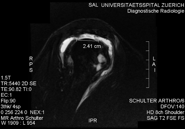
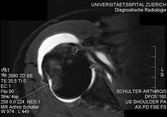
Figure 2: Partial Rupture
This is a partial rupture of one of the rotator cuff tendons. The red arrow indicates the rupture site.
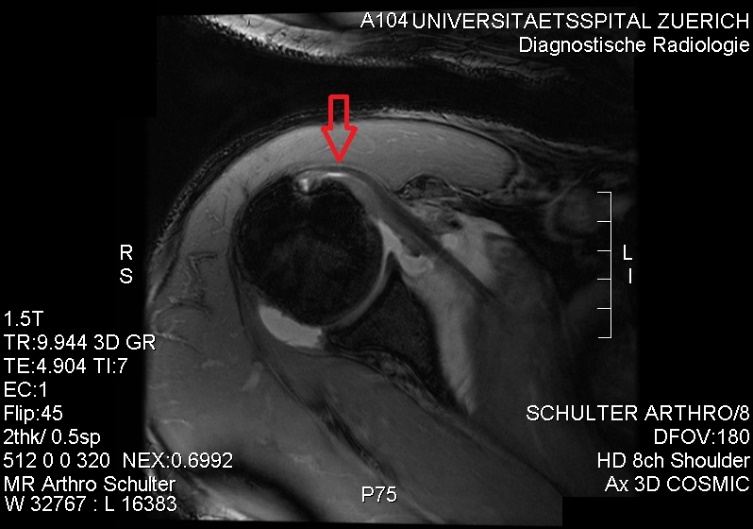
Figure 3: Complete Rotator Cuff Tear
This picture shows a complete rotator cuff tear. The arrow is pointing to the area of full thickness rupture through the tendon.
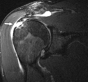
Figure 4: Shoulder Impingement
If rotator cuff tendons get “pinched” between the subacromial space and the acromion space, shoulder impingement occurs. Inflammation and swelling then follows and and leads to weakness, decreased range of motion, and pain in the shoulder area. MRI is helpful in differentiating this syndrome from an actual tear.
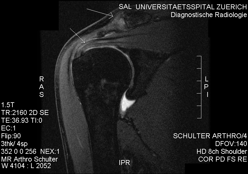
Figure 5: Edema/Rotator Cuff Tendon
This MRI shows actual swelling and inflammation near one of the rotator cuff tendons.
Treatment for Rotator Cuff Tear
A rotator cuff tear MRI can tell doctors what needs to be done for treatment. They can includesurgery, physical therapy, medications, or a combination of all three. The treatment plan depends on the extent of the injury. These include:
Non-Surgical
For injuries that do not require surgery, the doctor may try the following:
- Anti-inflammatory medications
- Steroid injections or oral steroids
- Physical therapy
- Modified duties at work
Surgical
If the doctor needs to do a surgical repair of the rotator cuff, there are a few different options for surgical repair. The doctor will either “debride” any damaged tissue from the joint or perform acromioplasty to remove some of the ligament tissue and bone to increase the range-of-motion to the shoulder. This can be done with one of these surgical techniques:
- Arthroscopic – The surgeon makes a few tiny incisions and inserts a scope with a camera. They can insert any needed tools to perform repairs.
- Open Surgery – For more severe tears, the doctor may need to make a large cut and completely repair the damaged tendon with regular surgical instruments. This type of surgery takes a little longer to recover than arthroscopy.
- Mini Surgery – This is a combination of both arthroscopy and open surgery, but the incision doesn’t have to be as large.